





G GCSE Apps

GCSE Science

B2 Organisation

11 subtopics in this section

Organisation and the digestive system

Definition

Levels of organisation, smallest to largest: cell → tissue → organ → organ system → organism.

A tissue is a group of similar cells doing one job. An organ (e.g. the stomach) is several tissues working together. An organ system is several organs working together.

The digestive system breaks down large insoluble food into small soluble molecules and absorbs them.

Method

Enzymes digest food into small soluble molecules:

Carbohydrases (e.g. amylase) break carbohydrates into simple sugars.

Proteases break proteins into amino acids.

Lipases break lipids (fats) into glycerol and fatty acids.

Products are used to build new molecules, and glucose is used in respiration.

Bile (made in the liver, stored in the gall bladder) is an alkaline liquid. It neutralises stomach acid and emulsifies fats into droplets, increasing their surface area so lipases work faster. Bile is not an enzyme.

Diagram
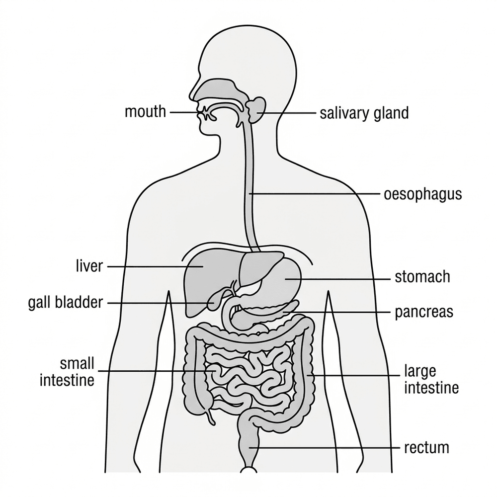
bio digestive system

Note

The human digestive system. Food passes down the oesophagus to the stomach, then the small intestine and large intestine.

Example

Describe what happens to the starch in a piece of bread as it passes through the digestive system.

Solution

In the mouth, amylase from the salivary glands starts breaking starch down into sugars.

In the small intestine, amylase from the pancreas and the small intestine finishes the job.

The sugars are small and soluble, so they are absorbed through the wall of the small intestine into the blood.

Cells use the glucose for respiration or to build new carbohydrates.

Tips/hints

Bile does not break chemical bonds.

The liver makes bile; the gall bladder only stores it.

The small intestine absorbs digested food; the large intestine absorbs water.
Enzymes

Definition

Enzymes are biological catalysts. They are large protein molecules that speed up reactions in living organisms without being used up.

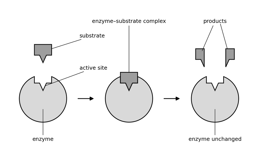
Each enzyme has an active site with a particular shape. In the lock and key model, only a substrate with a complementary shape fits, so each enzyme is specific to one reaction.

Diagram

bio enzyme lock and key

Note

The substrate fits the active site exactly, forming the enzyme–substrate complex. The products then leave and the enzyme is unchanged, ready to be used again.

Method

Enzymes work fastest at their optimum temperature (about 37 °C for human enzymes) and optimum pH.

At low temperatures the rate is low because the molecules collide less often. Above the optimum, or at extreme pH, the active site changes shape, the substrate no longer fits and the enzyme is denatured.

Rate = amount of product formed (or substrate used) ÷ time taken, e.g. in cm3/s. A rate can also be found as 1 ÷ time taken, in /s.

Example

An enzyme reaction is timed at four temperatures: 20 °C takes 80 s, 30 °C takes 40 s, 40 °C takes 25 s, and at 60 °C the reaction never finishes. Find the rate at 40 °C (rate = 1 ÷ time) and explain the 60 °C result.

Solution

Rate at 40 °C = 1 ÷ 25 = 0.040 /s – the fastest of the four, so the optimum is near 40 °C.

At 60 °C the active site has changed shape, so the substrate no longer fits: the enzyme is denatured and the reaction stops.

Tips/hints

Enzymes are molecules, not living things: say 'denatured', never 'killed'.

Denaturing is permanent – cooling or changing the pH back does not restore the active site.

When calculating a rate, check whether minutes must be converted to seconds.

The heart and blood vessels

Definition

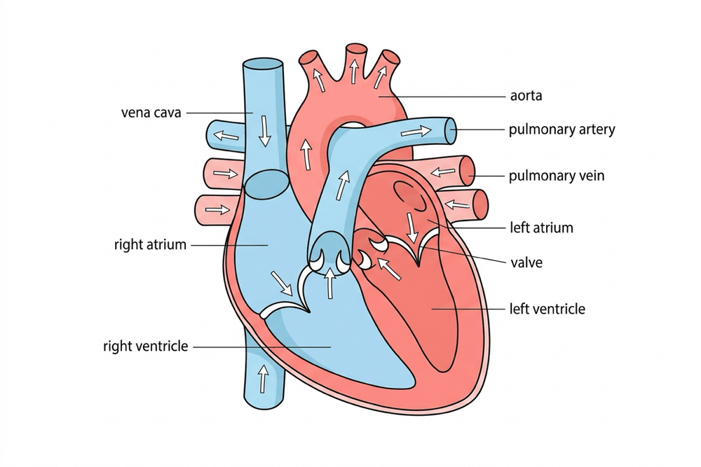
The heart is a muscular organ that pumps blood in a double circulatory system: blood passes through the heart twice in each complete circuit. The right side pumps blood to the lungs; the left side pumps it to the rest of the body.

Method

Blood from the body enters the right atrium through the vena cava, passes to the right ventricle and is pumped to the lungs in the pulmonary artery.

Oxygenated blood returns in the pulmonary vein to the left atrium, then the left ventricle pumps it to the body through the aorta. Its wall is thicker because it pumps blood all round the body.

Valves stop backflow. The coronary arteries supply the heart muscle. Cells in the right atrium act as the natural pacemaker; an artificial pacemaker corrects an irregular rhythm.

In the lungs, air passes down the trachea and bronchi to the alveoli, which have a huge surface area, thin walls and a rich capillary network.

Diagram

bio heart structure

Note

Drawn as if you face the person, so the heart's left side is on the right of the picture. Arrows show the direction of blood flow.

Diagram

bio blood vessels

Note

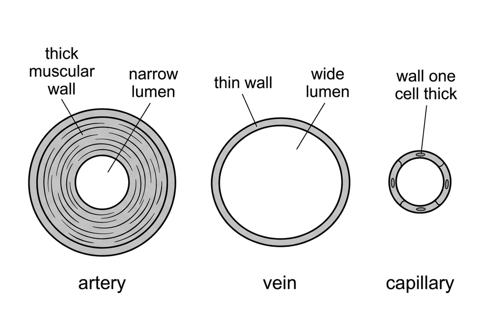
An artery has a thick muscular wall and a narrow lumen; a vein has a thin wall and a wide lumen; a capillary wall is one cell thick.

Example

A heart beats 70 times a minute and pumps 75 ml of blood per beat. How many litres does it pump each minute?

Solution

70 × 75 = 5250 ml per minute

5250 ÷ 1000 = 5.25 litres per minute

Tips/hints

The pulmonary artery carries deoxygenated blood: arteries carry blood away from the heart, whatever its oxygen content.

The heart does not oxygenate blood – the lungs do.

In flow-rate questions, 1 litre = 1000 ml and 1 minute = 60 s.

Blood

Definition

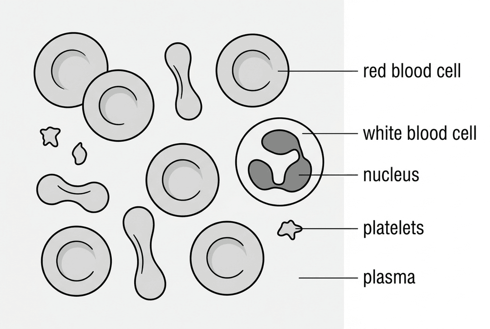
Blood is a tissue consisting of a liquid called plasma, which has red blood cells, white blood cells and platelets suspended in it.

Plasma is a pale yellow liquid that transports dissolved substances around the body. These include glucose, amino acids, carbon dioxide, urea and hormones, as well as the blood cells themselves.

Diagram

bio blood cells

Note

Red blood cells (no nucleus, some seen side-on), a larger white blood cell with a lobed nucleus and tiny platelets, all carried in plasma.

Example

A student looks at a blood smear under a microscope. They see a cell with no nucleus and a biconcave disc shape. What is this cell, and how is it adapted for its function?

Solution

It is a red blood cell. It carries oxygen from the lungs to the body cells.

Having no nucleus means there is more room for haemoglobin, which binds to oxygen. The biconcave disc shape gives it a large surface area to volume ratio, so oxygen can diffuse in and out quickly.

Tips/hints

Red blood cells do not have a nucleus, but white blood cells do.

White blood cells are part of the immune system. Some engulf pathogens (phagocytosis), while others make antibodies or antitoxins.

Platelets are tiny fragments of cells with no nucleus, not whole cells. They help the blood to clot at a wound, stopping blood loss and keeping pathogens out.

Haemoglobin is found inside red blood cells, not floating freely in the plasma.

Coronary heart disease

Definition

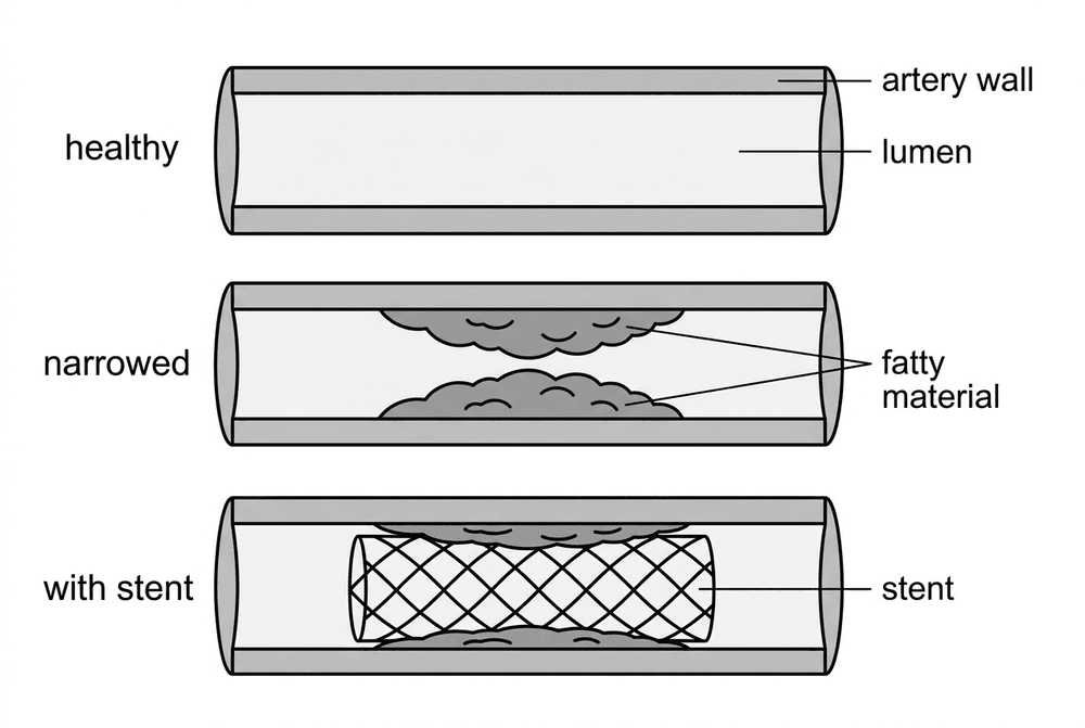
Coronary heart disease (CHD) is a non-communicable disease. Layers of fatty material build up inside the coronary arteries, narrowing them.

This reduces blood flow to the heart muscle, so it receives less oxygen and glucose for respiration. This can cause chest pain or a heart attack.

Diagram

bio coronary artery stent

Note

A healthy artery has a wide lumen. Fatty material on the artery wall narrows the lumen and reduces blood flow. A stent holds the artery open again.

Method

Stents: wire mesh tubes inserted to keep coronary arteries open. They work immediately and recovery is quick, but there is a risk of surgery complications or blood clots.

Statins: drugs that lower blood cholesterol to slow the rate of fatty deposit build-up. They prevent CHD without surgery, but must be taken long-term, have side effects, and do not remove existing deposits.

Heart valves: faulty valves can leak (causing backward flow) or not open fully, making the heart inefficient. They are replaced with biological valves (wear out sooner) or mechanical valves (need lifelong anti-clotting drugs).

Heart failure: treated with a donor transplant (risk of rejection, shortage of donors) or an artificial heart temporarily to keep the patient alive.

Example

A patient has a blood cholesterol concentration of 7.5 mmol/dm3. After six months of statins it is 6.0 mmol/dm3. What is the percentage decrease?

Solution

Decrease = 7.5 − 6.0 = 1.5 mmol/dm3

Percentage decrease = decrease ÷ original value × 100 = 1.5 ÷ 7.5 × 100 = 20%

Tips/hints

Coronary heart disease affects the coronary arteries that supply the heart muscle, not the blood travelling through the heart chambers.

Statins slow down new fatty deposits; they do not dissolve existing fat.

A stent treats narrowed arteries; a replacement valve treats a leaky or stiff heart valve.

Health issues and lifestyle

Definition

Health is the state of physical and mental well-being, not just the absence of disease. Diseases can be communicable (caused by pathogens and passed on) or non-communicable (cannot be passed on).

Different diseases often interact. A defective immune system makes infectious diseases more likely. Severe physical illness can trigger mental health issues like depression. Some viruses living in cells can trigger cancers, and immune reactions can trigger allergies.

Diagram

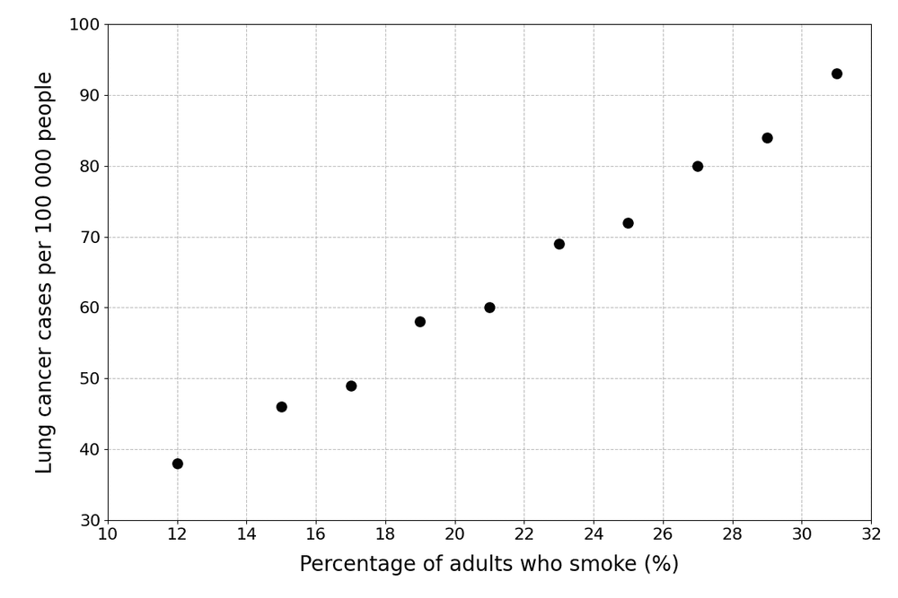
bio risk factor scatter

Note

This scatter diagram shows a positive correlation between smoking and lung cancer rates. However, correlation alone does not prove that smoking causes the cancer (other evidence is needed to prove the causal mechanism).

Example

Use the scatter diagram. Describe the relationship it shows, and explain whether it proves that smoking causes lung cancer.

Solution

As the percentage of adults who smoke rises from 12% to 31%, lung cancer cases rise from 38 to 93 per 100 000: a positive correlation.

The towns may differ in other ways (for example air pollution or age), so a correlation alone does not prove cause. The proof comes from a causal mechanism: chemicals in smoke damage the DNA of lung cells.

Tips/hints

A risk factor is linked to a higher rate of a disease; it does not mean a person will certainly get it.

Causal mechanisms are proven for some risk factors, such as smoking and lung cancer, and alcohol and liver damage.

Compare rates per 100 000 people, not raw numbers of cases, when populations differ in size.

Non-communicable diseases have human costs (illness, early death) and financial costs (NHS treatment, lost earnings).

Cancer

Definition

Cancer is caused by changes in cells (to their DNA) that make them grow and divide in an uncontrolled way, forming a mass of abnormal cells called a tumour.

Method

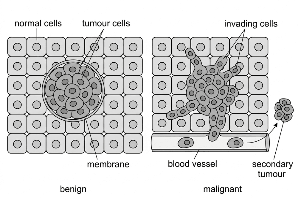
Benign tumours are growths of abnormal cells that stay in one place, usually surrounded by a membrane. They do not invade other tissues. They are not cancers, but they can still be dangerous if they press on organs, such as the brain.

Malignant tumours are cancers. Their cells can invade nearby healthy tissues. Cells can also break away and travel in the blood or lymphatic system to other parts of the body, where they form secondary tumours.

Diagram

bio benign malignant tumour

Note

A benign tumour stays inside its membrane. Malignant tumour cells invade the normal cells around them, enter a blood vessel and form a secondary tumour elsewhere.

Example

What lifestyle and genetic risk factors can increase the chance of developing cancer?

Solution

Lifestyle factors: Smoking is a major risk factor for lung cancer. Obesity, excessive alcohol intake, and UV exposure (from sunbathing or sunbeds, which increases skin cancer risk) are also linked to cancer.

Genetic factors: Inheriting certain faulty alleles, such as the BRCA genes, can increase the risk of developing breast cancer.

Carcinogens: Factors that cause cancer are called carcinogens. Ionising radiation (such as X-rays and gamma rays) is a carcinogen because it can damage DNA.

Tips/hints

Remember that not all tumours are cancer; only malignant tumours are classified as cancer.

Benign does not always mean harmless! A benign tumour can still cause severe damage by putting pressure on vital organs.

Cancer is not infectious. You cannot catch it from another person.

Secondary tumours are formed from malignant cells that have travelled in the blood, so they are the same type of cancer as the original tumour.

Plant tissues

Definition

A leaf is an organ made of several tissues working together. Its main role is to absorb light for photosynthesis to make food for the plant.

Method

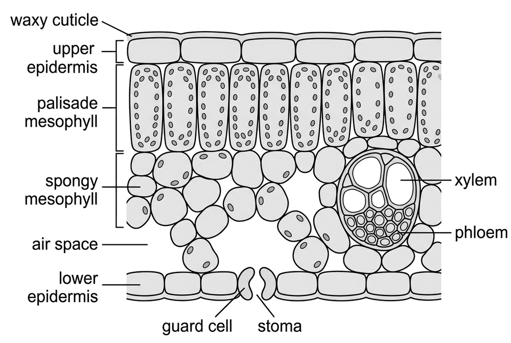
The epidermis (upper and lower) is a thin, transparent layer protecting the leaf. The upper epidermis has a waxy cuticle to reduce water loss.

The palisade mesophyll contains tall cells packed with chloroplasts. It is near the top to absorb maximum light for photosynthesis.

The spongy mesophyll has loosely packed cells with large air spaces, allowing gases (carbon dioxide, oxygen) to diffuse easily.

Veins contain xylem (carries water and minerals to the leaf) and phloem (carries sugars away).

Guard cells around stomata open and close to control gas exchange and water loss.

Meristem tissue at the tips of shoots and roots contains cells that divide and differentiate.

Diagram

bio leaf cross section

Note

A transverse section of a leaf. The palisade mesophyll lies just under the upper epidermis to absorb light; the spongy mesophyll has air spaces leading to the stoma between two guard cells; the vein contains xylem above phloem.

Example

A student counts the stomata on the lower surface of a leaf in three fields of view, each 0.5 mm2: 12, 15 and 18. What is the mean number of stomata per mm2?

Solution

Mean per field of view = (12 + 15 + 18) ÷ 3 = 45 ÷ 3 = 15

Stomata per mm2 = 15 ÷ 0.5 = 30 stomata per mm2

Tips/hints

The palisade mesophyll is the main site of photosynthesis, not the epidermis.

Stomata are mainly on the lower surface to reduce water loss.

Remember: xylem carries water; phloem carries food ('flow').

A leaf is an organ, not a tissue, as it is made of multiple tissues.

Transpiration and translocation

Definition

The plant transport system consists of roots, stem and leaves.

Xylem tissue is made of hollow tubes of dead cells with no end walls, strengthened by lignin. It carries water and mineral ions from the roots up to the leaves (the transpiration stream).

Phloem tissue is tubes of elongated living cells with pores in their end walls. It moves dissolved sugars made in the leaves to the rest of the plant for use or storage. This is translocation.

Method

Transpiration is the loss of water vapour from the leaves. Water evaporates inside the leaf and diffuses out through the stomata, pulling more water up the xylem.

Water enters the roots through root hair cells and is carried up the stem in the xylem to replace the water lost from the leaves.

Diagram

bio potometer

Note

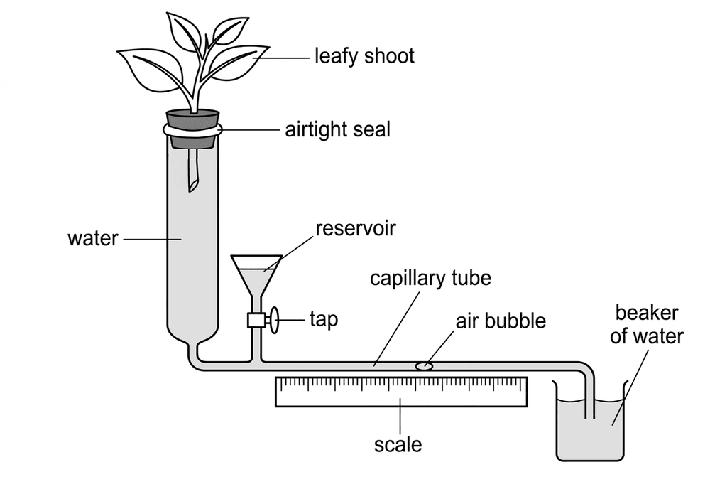
A bubble potometer measuring water uptake by a leafy shoot. The capillary tube contains an air bubble. The reservoir and tap reset the bubble.

Example

A student uses a potometer to measure water uptake. The bubble moves 54 mm in 15 minutes. Calculate the rate in mm/min.

Solution

Rate of water uptake = distance moved ÷ time

Rate = 54 ÷ 15 = 3.6 mm/min.

Tips/hints

Transpiration increases with higher temperature (faster evaporation), lower humidity (steeper gradient), more air movement and higher light intensity (stomata open).

Guard cells control stomata, closing them in the dark or when short of water to reduce water loss.

A potometer measures water uptake. This slightly overestimates transpiration because some water is used in photosynthesis.

Required practical: Food tests

Overview

Food tests use chemical reagents to find out which nutrients a food contains. They show whether a nutrient is present, not the exact amount.

Method

1. Grind the food with a pestle and mortar.

2. Add distilled water and stir to dissolve the nutrients.

3. Filter the mixture (or let solids settle) to get a clear food solution.

4. Test the solution using the correct reagent for each nutrient.

Knowledge Required

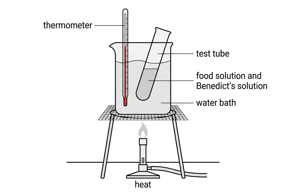
Reducing sugars (e.g. glucose): Add Benedict's solution (blue) and heat in a water bath at 75–80 °C for 5 minutes. A green, yellow or brick-red precipitate is a positive result.

Starch: Add iodine solution (orange-brown). A blue-black colour is a positive result.

Protein: Add Biuret reagent (blue). A purple or lilac colour is a positive result.

Lipids (fats): Use the ethanol emulsion test. Shake the food with ethanol, then pour it into water. A cloudy white emulsion is a positive result.

Note

Benedict's solution and Biuret reagent are irritants (Biuret contains corrosive sodium hydroxide): wear eye protection.

Ethanol is highly flammable: keep it away from flames, and heat Benedict's in a water bath, not over a flame.

Diagram

bio benedicts water bath

Note

A water bath allows you to safely control the temperature when heating Benedict's solution.

Example

A student tests an unknown drink. It stays blue with Benedict's solution and stays orange-brown with iodine solution, but turns purple with Biuret reagent. Which nutrients are present?

Solution

The drink contains protein (shown by the purple Biuret result).

It does not contain reducing sugars or starch (both gave negative results).

Tips/hints

Table sugar (sucrose) is a non-reducing sugar, so it gives a negative result (stays blue) with Benedict's.

Benedict's test is semi-quantitative: a brick-red result means more reducing sugar than a green result.

Use a control tube of distilled water to show the negative colour for comparison.

Required practical: Enzymes and pH

Overview

This practical investigates how pH affects the rate at which the enzyme amylase digests starch.

Method

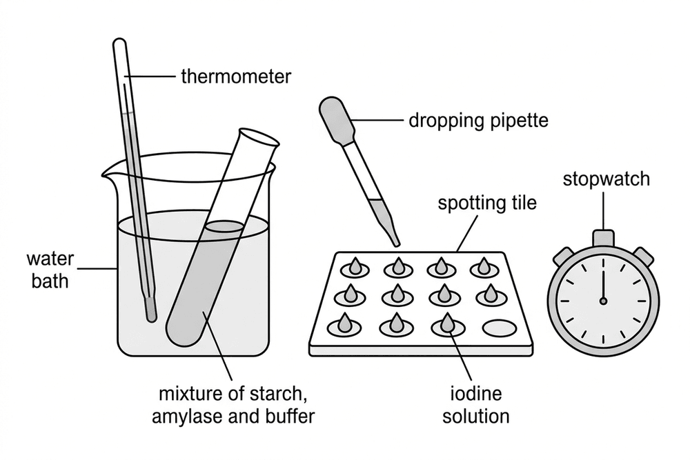
1. Put one drop of iodine solution in each well of a spotting tile.

2. Place test tubes of starch, amylase and a pH buffer in a water bath (e.g. 35 °C) for a few minutes to reach the same temperature.

3. Mix the amylase, starch and buffer and start a stopwatch.

4. Every 30 seconds, take a drop of the mixture with a pipette and add it to a fresh well of iodine.

5. Record the time when the iodine stays orange-brown (no blue-black appears). This means all starch is digested.

6. Repeat with different pH buffers (e.g. 4, 5, 6, 7, 8), and repeat for each pH.

Knowledge Required

Variables: Independent is pH (using a buffer). Dependent is time for starch to be digested. Control variables include temperature (using a water bath), volumes and concentrations of starch, amylase and buffer, sampling interval and iodine volume.

Note

Hazards: Iodine is an irritant and stains; wear eye protection. Amylase can be an allergen; avoid skin contact. Take care with the hot water bath.

Diagram

bio amylase ph apparatus

Note

Apparatus for investigating the effect of pH on amylase. A water bath is used to control the temperature.

Tips/hints

Sampling only every 30 s means the end point is known only to ±30 s. Sample more often (e.g. every 10 s) for better resolution.

Judging the colour change is subjective; using a colour reference or a colorimeter improves this.

Rate is calculated as rate = 1 ÷ time. A shorter time means a faster rate.

If the iodine stays blue-black for the whole experiment, it means the starch was not digested because the enzyme denatured.

Test yourself on this topic

Try the free quiz with worked solutions.

Start quiz →

Get GCSE Science

Full notes, full question bank, offline.