



G GCSE Apps

GCSE Science

B1 Cell biology

11 subtopics in this section

Eukaryotes and prokaryotes

Definition

Eukaryotic cells (animal, plant, fungal and protist cells) have a cell membrane, cytoplasm and genetic material inside a nucleus.

Prokaryotic cells (bacteria) are much smaller. They have cytoplasm and a cell membrane surrounded by a cell wall (not made of cellulose). Their genetic material is a single loop of DNA, not enclosed in a nucleus, and there may be small extra rings of DNA called plasmids.

Method

Cell sizes use metric prefixes: centi (c) = 10−2, milli (m) = 10−3, micro (μ) = 10−6, nano (n) = 10−9.

1 mm = 1000 μm and 1 μm = 1000 nm. To go to a smaller unit multiply by 1000; to go to a larger unit divide by 1000.

Typical sizes: a bacterium is about 1–5 μm long, an animal cell about 10–30 μm and a plant cell about 10–100 μm.

An order of magnitude is a factor of 10. Something 100 times (102) larger is two orders of magnitude larger.

Diagram
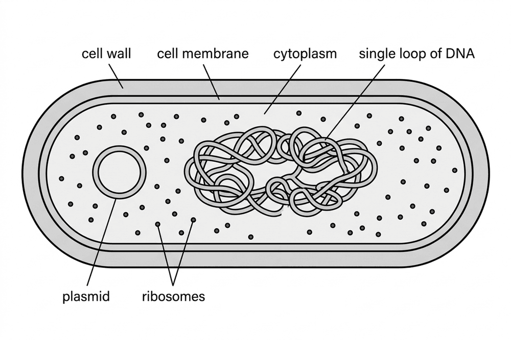
bio bacterial cell

Note

A bacterial (prokaryotic) cell. Its genetic material is a single loop of DNA, with a small ring of DNA called a plasmid, and there is no nucleus. There are no mitochondria or chloroplasts.

Example

A plant cell is 40 μm long and a bacterium is 4 μm long. How many orders of magnitude larger is the plant cell?

Solution

Both are in μm, so divide: 40 ÷ 4 = 10.

10 = 101, so the plant cell is one order of magnitude larger.

Tips/hints

Put both sizes in the same unit before you compare them.

Bacteria do have a cell wall, but it is not made of cellulose.

Plasmids are extra DNA. The main genetic material of a bacterium is the single DNA loop.

Prokaryotic cells have no nucleus and no mitochondria, but they do have ribosomes.
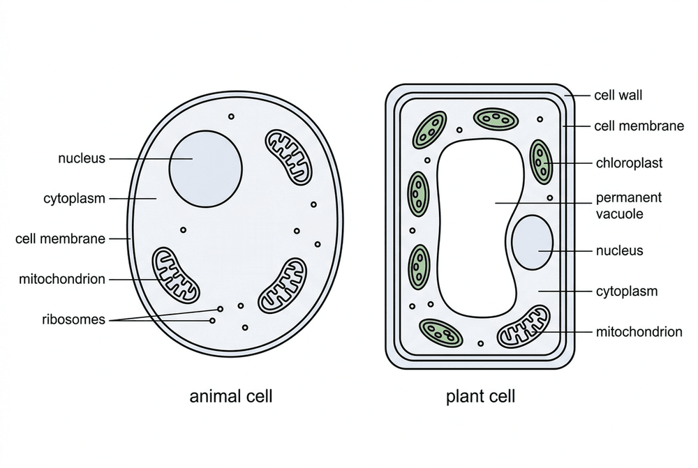
Animal and plant cells

Definition

Most animal cells have a nucleus, cytoplasm, a cell membrane, mitochondria and ribosomes. Plant cells have these too, and often also chloroplasts, a permanent vacuole and a cellulose cell wall (algal cells also have a cellulose wall).

Method

The function of each structure relates to what the cell does. The nucleus holds the genetic material that controls the cell's activities. The cytoplasm is a jelly-like substance where most chemical reactions take place. The cell membrane controls which substances enter and leave the cell.

Mitochondria are where aerobic respiration happens, releasing energy for the cell. Ribosomes are where proteins are made (protein synthesis).

In plant cells, the cell wall strengthens and supports the cell. Chloroplasts contain chlorophyll, which absorbs light for photosynthesis. The permanent vacuole keeps the cell firm (turgid) and so supports the plant.

Diagram

bio animal and plant cells

Note

A typical animal cell and plant cell. The plant cell also has a cell wall, chloroplasts and a large permanent vacuole; both cells have a nucleus, cytoplasm, a cell membrane and mitochondria.

Example

A plant cell is 80 μm long and its nucleus is about 10 μm across. Estimate how many times longer the cell is than the width of the nucleus.

Solution

Both lengths are in μm, so divide: 80 ÷ 10 = 8.

The cell is about 8 times longer than the nucleus is wide.

Tips/hints

Plant cells have mitochondria as well as chloroplasts: they respire all the time to release energy.

The cell wall is freely permeable and gives strength. It is the cell membrane that controls what enters and leaves the cell.

Not every plant cell has chloroplasts. Root cells receive no light, so they have none.

Link structure to function: lots of energy needed means many mitochondria; lots of protein (such as enzymes) made means many ribosomes.

Cell specialisation and differentiation

Definition

A specialised cell has a structure that suits it to one particular job.

Differentiation is the process by which a cell becomes specialised, gaining the sub-cellular structures it needs. Most animal cells differentiate early in development; in mature animals, cell division is mainly for repair and replacement. Many plant cells can differentiate throughout the plant's life.

Method

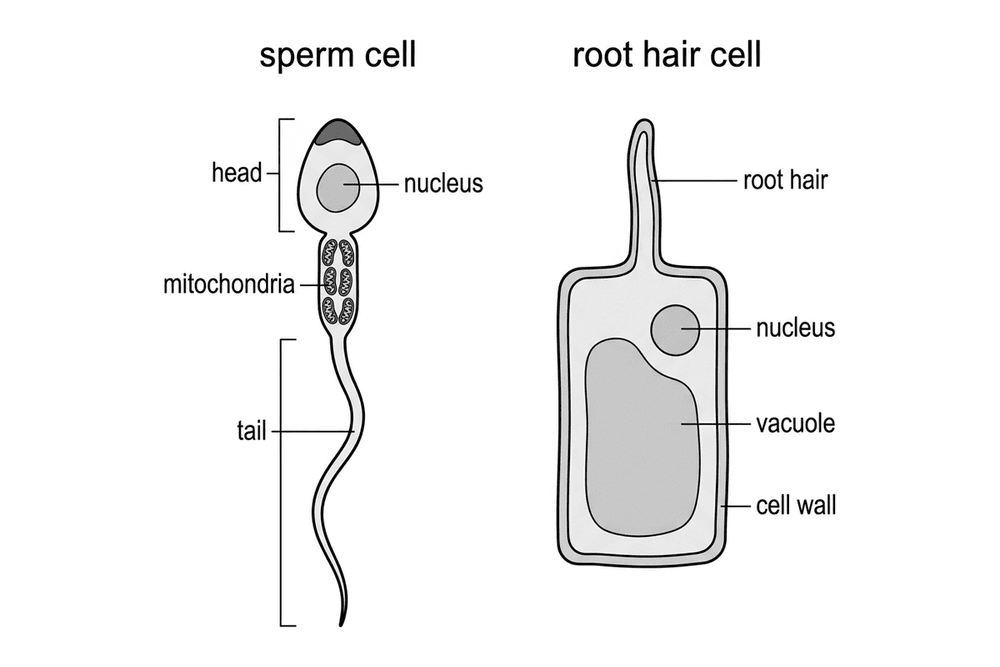
Sperm cell: tail to swim; many mitochondria release energy; enzymes in the head digest the egg's outer layer; nucleus with half the genetic material.

Nerve cell: long axon carries impulses a long way; branched endings connect to other cells.

Muscle cell: protein fibres slide to contract; many mitochondria.

Root hair cell: long projection gives a large surface area for absorbing water and mineral ions; no chloroplasts.

Xylem: dead, hollow tubes with no end walls, strengthened by lignin; carry water and mineral ions upwards.

Phloem: living cells with perforated end walls and companion cells; carry dissolved sugars up and down.

Diagram

bio specialised cells

Note

The sperm cell's tail and mitochondria help it swim. The root hair cell's long root hair gives a large surface area for absorbing water and mineral ions.

Example

A cell lining the small intestine has many tiny folds on its outer surface and contains many mitochondria. Suggest how these features help it absorb digested food.

Solution

The folds give a large surface area, so more food molecules can be absorbed at once.

Many mitochondria release lots of energy by respiration, which the cell can use to absorb food molecules by active transport.

Tips/hints

For an unfamiliar cell, link each feature to the job: shape or surface area, number of mitochondria, and missing or extra structures.

Xylem cells are dead; phloem cells are living.

Root hair cells have no chloroplasts because they are underground.

Microscopy

Definition

Light microscopes use light and lenses to form an image of a specimen. They can view living specimens in natural colours, but their magnification (up to roughly ×2000) and resolution (about 0.2 μm) are limited.

Electron microscopes use a beam of electrons instead of light. They provide a much higher magnification (up to millions of times) and a much higher resolving power. This allows biologists to see finer detail and understand sub-cellular structures like ribosomes and mitochondria inner membranes. However, specimens must be viewed in a vacuum, so they are dead.

Resolution (or resolving power) is the smallest distance between two points that can still be seen as separate. Better resolution gives a sharper, more detailed image.

Magnification is how many times larger the image is than the real object.

Method

magnification = size of image ÷ size of real object

Rearranged: size of real object = size of image ÷ magnification, and size of image = magnification × size of real object.

Both sizes must be in the same unit. To convert millimetres (mm) to micrometres (μm), multiply by 1000.

Magnification has no unit. It is written with a times sign (e.g. ×400).

Example

An electron micrograph shows a mitochondrion 15 mm long at a magnification of ×6000. What is the real length of the mitochondrion in μm?

Solution

Real size = size of image ÷ magnification = 15 ÷ 6000 = 0.0025 mm

Convert to μm: 0.0025 × 1000 = 2.5 μm

Tips/hints

Always check that both sizes are in the same unit (e.g. both in mm or both in μm) before dividing.

Higher magnification without higher resolution just produces a larger, blurred image. Resolution is what provides the fine detail.

Electron microscopes cannot view living cells.

Chromosomes and mitosis

Definition

The nucleus contains chromosomes. Each chromosome is one long DNA molecule carrying many genes.

In body cells chromosomes are in pairs: human body cells have 23 pairs (46 chromosomes).

Mitosis is needed for growth and development, for repair, for replacing worn-out cells and for asexual reproduction.

Method

The cell cycle happens in three main stages.

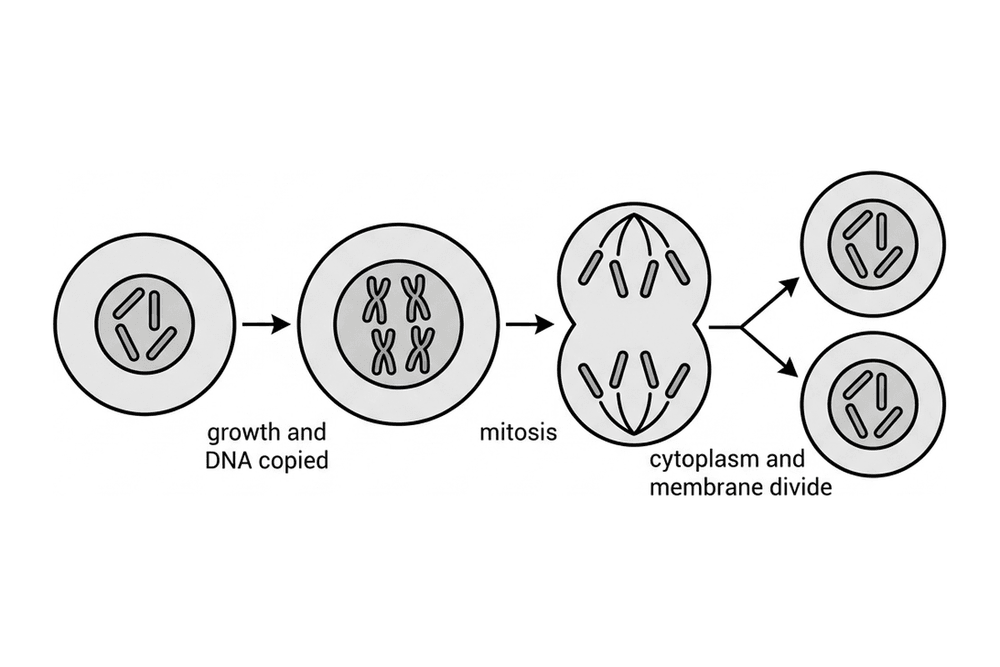
Stage 1: The cell grows and increases the number of sub-cellular structures such as ribosomes and mitochondria. The DNA replicates to form two copies of each chromosome.

Stage 2 (Mitosis): One set of chromosomes is pulled to each end of the cell and the nucleus divides.

Stage 3: The cytoplasm and cell membrane divide to form two genetically identical cells.

Diagram

bio cell cycle

Note

Stage 1: the cell grows and each of its 4 chromosomes is copied. Stage 2 (mitosis): one set of chromosomes is pulled to each end and the nucleus divides. Stage 3: the cytoplasm and cell membrane divide, giving two identical cells with 4 chromosomes each.

Example

A scraped knee heals over two weeks. Explain how mitosis repairs the skin.

Solution

Skin cells next to the wound go through the cell cycle: they grow, copy their DNA, then divide by mitosis.

Each division gives two genetically identical cells with the same number of chromosomes as the original cell.

Repeated divisions make enough new skin cells to replace the damaged ones.

Tips/hints

DNA is copied before mitosis starts, not during it.

Mitosis keeps the chromosome number the same: a body cell with 46 chromosomes gives two cells with 46 each. Halving the number happens in meiosis, which makes gametes.

To estimate the time spent in mitosis: (cells in mitosis ÷ total cells counted) × length of the cell cycle.

Stem cells

Definition

A stem cell is an undifferentiated cell. It can divide to make many more cells of the same type, and some of these can differentiate into other types of cell.

Embryonic stem cells can differentiate into most types of human cell. Adult stem cells, such as those in bone marrow, form fewer types of cell, including blood cells. Meristem tissue at plant root and shoot tips can form any type of plant cell throughout the plant's life.

Method

Possible treatments: stem cells may help treat conditions such as diabetes (new insulin-producing cells) and paralysis (new nerve cells). Bone marrow transplants already use adult stem cells.

Therapeutic cloning: an embryo is made with the same genes as the patient, so its stem cells are not rejected by the patient's body.

Plant cloning: meristem cells give many identical plants quickly and cheaply, to save rare species or grow crops with useful features such as disease resistance.

Example

Give one benefit, one risk and one ethical issue of using embryonic stem cells to treat type 1 diabetes.

Solution

Benefit: the stem cells could be made into insulin-producing cells, so the patient might not need insulin injections.

Risk: cells grown in the laboratory could pass on a viral infection.

Ethical issue: an embryo is destroyed to collect the stem cells, which some people object to on ethical or religious grounds.

Tips/hints

Adult stem cells cannot form every type of cell; embryonic stem cells can form most types.

Keep risks (such as infection) separate from ethical or religious objections (such as destroying embryos) when you evaluate.

Therapeutic cloning makes an embryo only as a source of stem cells, not to produce a baby.

Clones are genetically identical, so they have no variation to resist a new disease.

Diffusion

Definition

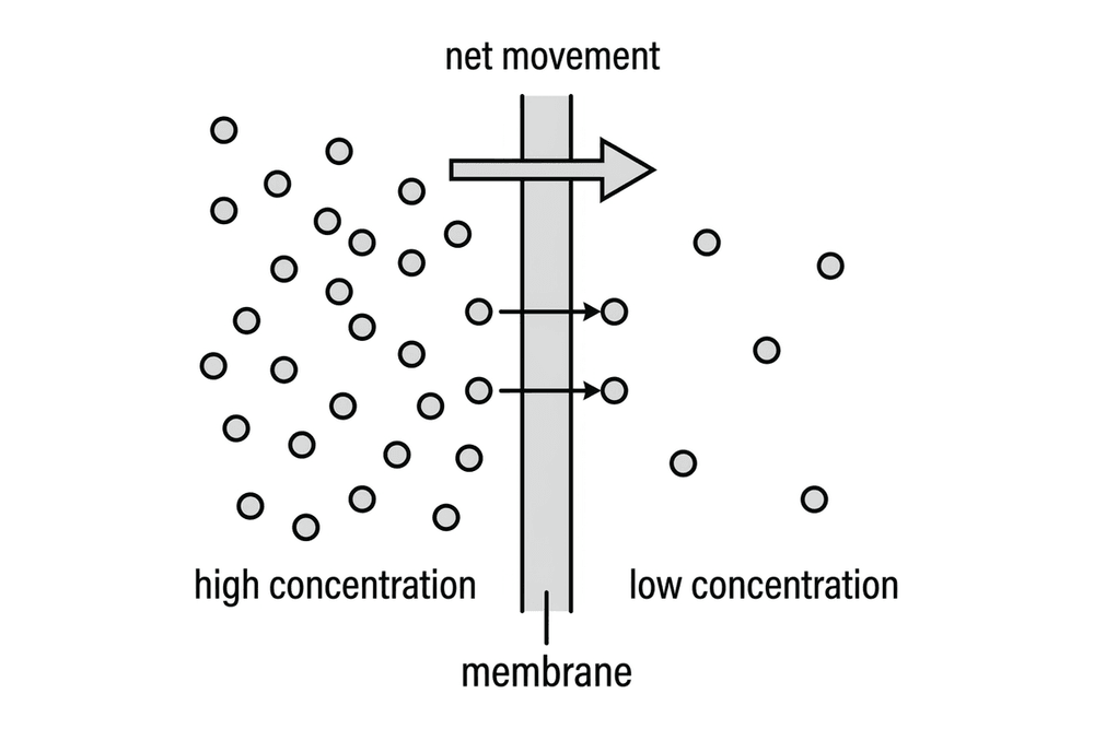
Diffusion is the spreading out of the particles of a gas, or of a substance in solution, giving a net movement from a region of higher concentration to a region of lower concentration.

Particles move randomly in all directions, but more move from the crowded side, so there is a net movement. Diffusion is passive: it needs no energy from respiration.

Examples: oxygen and carbon dioxide in gas exchange, and urea passing from cells into the blood plasma to be removed by the kidneys.

Method

Faster diffusion comes from a steeper concentration gradient, a higher temperature (particles move faster) and a larger membrane surface area.

Surface area to volume ratio: for a cube of side l, surface area = 6l2 and volume = l3. Write surface area : volume and simplify.

Exchange surfaces (villi in the small intestine, alveoli in the lungs, gill filaments, root hairs, leaves) work well because they have a large surface area and a thin membrane (short diffusion path). In animals, a good blood supply and ventilation keep the gradient steep.

Diagram

bio diffusion membrane

Note

Particles cross the membrane in both directions, but more cross from the high concentration side, so the net movement is from high to low concentration.

Example

Find the surface area to volume ratio of a cube with 2 cm sides.

Solution

Surface area = 6 × (2 × 2) = 24 cm2

Volume = 2 × 2 × 2 = 8 cm3

Ratio = 24 : 8 = 3 : 1

Tips/hints

Bigger objects have a smaller surface area to volume ratio. That is why large organisms need exchange surfaces and a transport system, while a single cell can rely on diffusion.

Give the ratio as surface area : volume, in its simplest form, and remember all six faces.

Osmosis

Definition

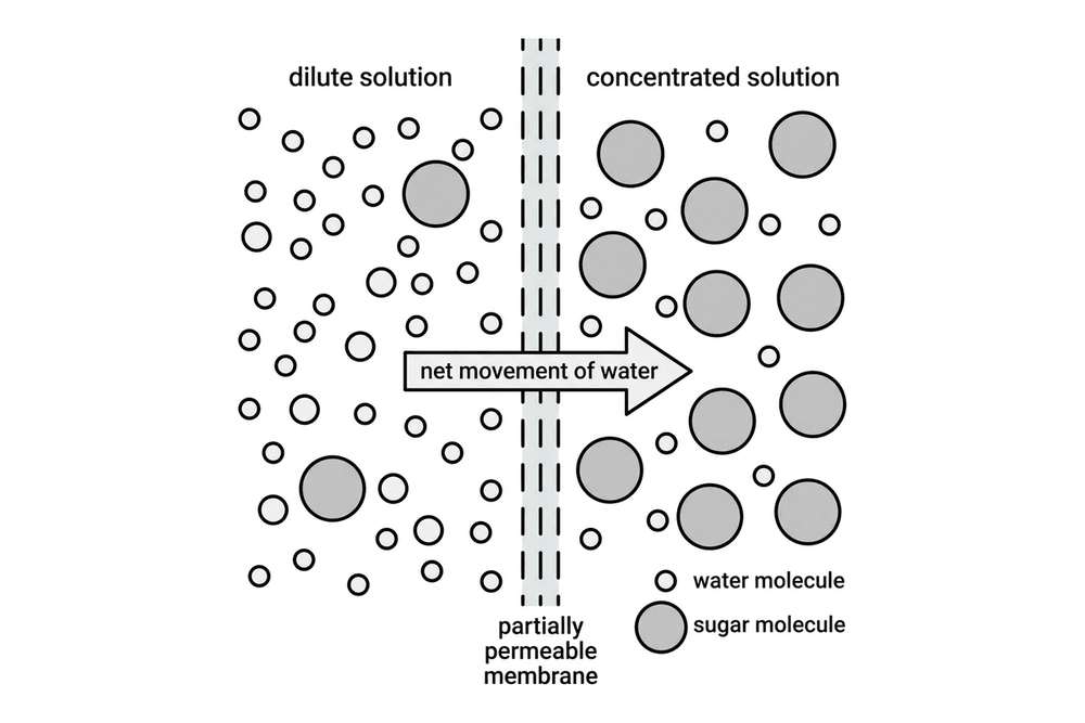
Osmosis is the diffusion of water from a dilute solution to a more concentrated solution through a partially permeable membrane.

A dilute solution has a high concentration of water and a low concentration of solute (like sugar). A concentrated solution has a low concentration of water and a high concentration of solute.

The partially permeable membrane lets small water molecules pass through, but blocks larger solute molecules. Osmosis is a passive process and does not require energy.

Method

To calculate percentage change in mass:

Percentage change = (final mass − initial mass) ÷ initial mass × 100

A negative sign means mass was lost; a positive sign means mass was gained.

Diagram

bio osmosis membrane

Note

Water molecules cross the membrane both ways, but more cross from the dilute side, so the net movement of water is into the concentrated solution. The sugar molecules are too large to pass through.

Example

A strip of rhubarb has a mass of 2.0 g. After an hour in pure water its mass is 2.3 g. What is the percentage change in mass?

Solution

Change in mass = 2.3 − 2.0 = +0.3 g

Percentage change = (0.3 ÷ 2.0) × 100 = +15%

The strip gained water by osmosis, because pure water is more dilute than the cell contents.

Tips/hints

Only water moves by osmosis, not the solute particles.

Animal cells (like red blood cells) placed in pure water will swell and may burst because they do not have a cell wall. Plant cells become turgid (firm) because their strong cellulose cell wall prevents bursting.

Plant cells in concentrated solutions lose water and become flaccid (soft). The cell membrane may pull away from the cell wall, which is called plasmolysis.

Always divide by the initial mass when calculating percentage change.

Active transport

Definition

Active transport moves substances from a more dilute solution to a more concentrated solution. This is against the concentration gradient.

Unlike diffusion and osmosis, it needs energy. The energy is released by respiration, so cells that do a lot of active transport contain many mitochondria.

Method

Plant roots: root hair cells absorb mineral ions, such as nitrate and magnesium, from very dilute soil water. The ions are already more concentrated inside the root, so diffusion cannot take them in. Plants need these ions for healthy growth.

Small intestine: sugar (glucose) is absorbed from the gut into the blood even when the blood already holds more sugar. The sugar is then used for respiration.

Comparing the three processes: diffusion moves particles down a concentration gradient with no energy; osmosis moves water from a dilute to a more concentrated solution across a partially permeable membrane with no energy; active transport moves substances against a concentration gradient using energy from respiration.

Example

Barley roots are kept in the same solution of potassium ions at 5 °C and at 25 °C. They take up potassium ions much more slowly at 5 °C. Suggest why.

Solution

The roots take up potassium ions by active transport, which needs energy from respiration.

Respiration is controlled by enzymes, which work more slowly at low temperatures.

At 5 °C less energy is released, so the ions are moved into the root cells more slowly.

Tips/hints

Say energy is released by respiration, not 'made' or 'created'.

Anything that slows respiration (little oxygen in waterlogged soil, a poison, low temperature) slows active transport. Diffusion and osmosis keep going because they are passive.

Osmosis is about water. Mineral ions and sugar enter cells by diffusion or active transport.

To decide which process is happening, compare the concentrations: moving into a higher concentration means active transport.

Required practical: Microscopy

Overview

Use a light microscope to observe, draw and label plant and animal cells, with a scale shown.

Method

1. Peel a thin layer of onion epidermis with forceps and lay it flat in a drop of water on a slide.

2. Add a drop of iodine solution to stain it. (For cheek cells: smear with a clean cotton bud and stain with methylene blue.)

3. Lower a cover slip at an angle with a mounted needle to avoid air bubbles.

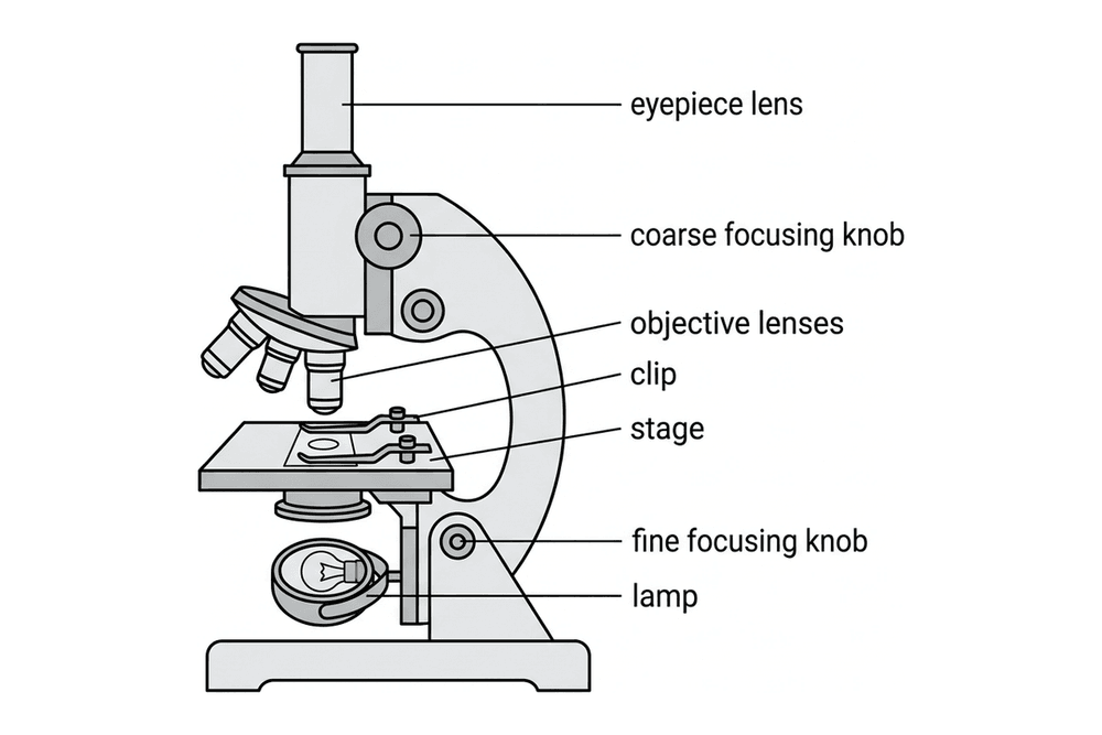
4. Clip the slide onto the stage and start with the lowest-power objective lens.

5. Focus with the coarse focusing knob, then sharpen with the fine focusing knob.

6. Change to a higher-power objective lens and refocus with the fine knob only.

7. Draw and label the cells, adding a title and magnification or scale bar.

Knowledge Required

An observation practical, so there are no variables to control.

Apparatus: light microscope, slides, cover slips, forceps, mounted needle, iodine solution, methylene blue.

Total magnification = eyepiece lens magnification × objective lens magnification.

Real size from a scale bar = measured length × scale value ÷ bar length.

Note

Hazards: iodine solution is an irritant, so wear eye protection. Take care with forceps and glass slides. Put used cotton buds straight into disinfectant.

Diagram

bio light microscope

Note

Light from the lamp passes through the slide, an objective lens and the eyepiece lens. The focusing knobs move the stage to focus.

Tips/hints

Drawings: sharp pencil, continuous lines, no shading, ruled label lines that do not cross.

Air bubbles look like black-edged circles; lower the cover slip at an angle to avoid them.

Onion cells show a cell wall, cytoplasm and nucleus but no chloroplasts. Ribosomes and mitochondria are too small to see clearly.

Estimate a cell's size: field of view diameter ÷ number of cells across it.

Required practical: Osmosis

Overview

Aim: find out how sugar or salt solution concentration affects plant tissue mass, and estimate the tissue's cell sap concentration.

Method

1. Use a cork borer to cut potato cylinders of the same diameter, and trim them to the same length with a scalpel on a white tile.

2. Blot each cylinder dry and weigh it on a balance.

3. Put each cylinder in a labelled boiling tube with the same volume of a different sugar solution (e.g. 0.0, 0.2, 0.4, 0.6, 0.8 and 1.0 mol/dm3).

4. Leave them for the same amount of time (e.g. 24 hours).

5. Remove them, blot them gently in the same way, and reweigh them.

6. Repeat the experiment at each concentration and calculate a mean.

Knowledge Required

Independent variable: concentration of the sugar solution.

Dependent variable: percentage change in mass.

Control variables: cylinder size and surface area, potato type, volume of solution, time left in solution, and temperature.

Note

Hazards: the cork borer and scalpel are sharp. Cut on a tile, away from your body.

Diagram

bio osmosis potato graph

Note

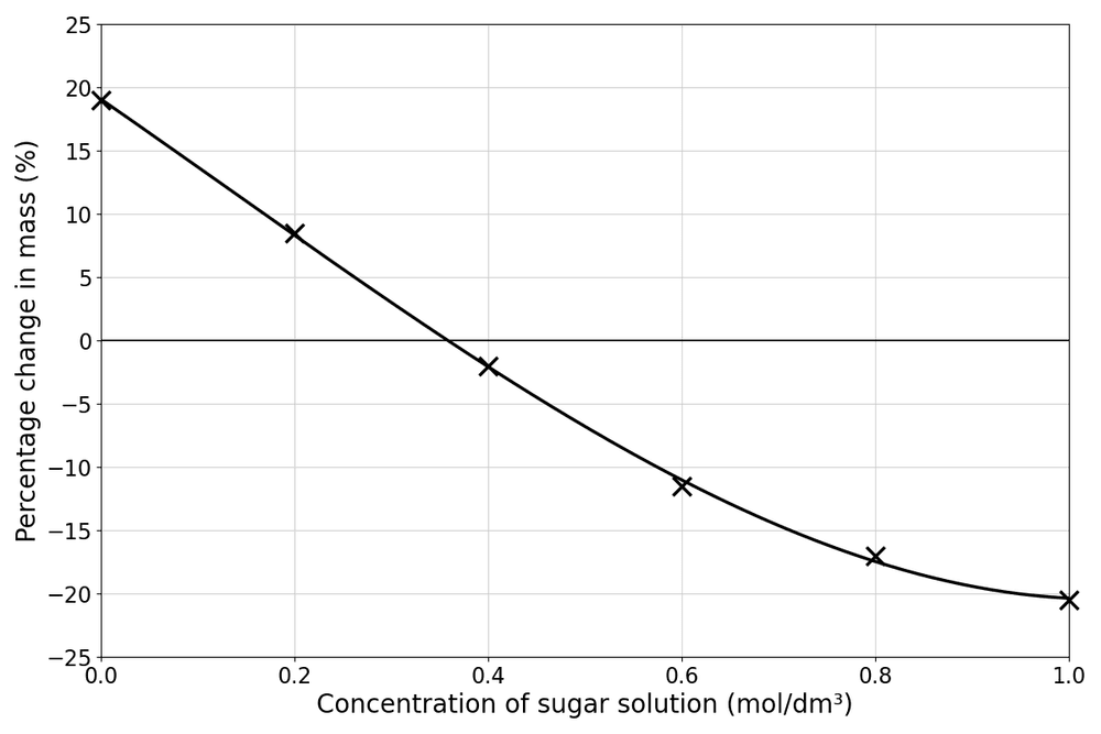
Example results. The line of best fit crosses zero change at about 0.36 mol/dm3: at this concentration there is no net osmosis, so it is the estimated concentration of the potato cell sap.

Tips/hints

Percentage change = (final mass − initial mass) ÷ initial mass × 100. It is used because the cylinders start at slightly different masses.

A gain in mass means the solution is more dilute than the cell sap; a loss means it is more concentrated.

Blot every cylinder in the same way: solution left on the surface adds mass.

Leave anomalous results out of the mean, and use smaller concentration steps near the crossing point for a better estimate.

Test yourself on this topic

Try the free quiz with worked solutions.

Start quiz →

Get GCSE Science

Full notes, full question bank, offline.